

|
|
|
|
|
|
|
|
|
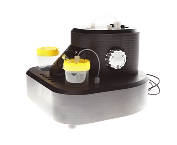
BioScan atomic weighter
The unique multifunctional analyzer of chemical and biological substances.
System advantages:
-
Tension measurement of thin films
-
Cell working volume 200 μ
-
The sensitivity of the cantilever deflection detection system is 1 nm
-
Using 4 cantilevers for direct multivariate analysis
-
Additional monitoring of the measuring cell using an optical camera
-
Convenient sample entry system
-
Record detection limit: 10-14 mol / liter
-
The device connects to the computer via a USB cable and does not require additional power
-
Intuitive program interface
-
Analysis of the kinetics of the formation of monolayers and surface chemical reactions
Universal cantilever sensor
The technology of cantilever sensors theoretically allows you to measure mass with an accuracy of 10-18 grams, which is comparable to the mass of a single protein molecule. In order to put such a sensitive system into practice, we developed a unique BioScan device for registering supersmall quantities of a substance in gases and liquids. It combines the functions of biological and chemical nanosensors. Main advantages: record sensitivity by weight, minimal amounts of reagent, multifunctionality, resistance to acts of nonspecific binding, compactness.
How it works
The analyzed mixture is introduced into the cell in which cantilevers are installed, and a laser beam is directed at them. After reflection from the cantilevers, the mirror system directs the beam to the detector - the program analyzes the camera matrix, the image it receives. Chemical reactions and the processes of adsorption and desorption on the surface of the cantilever cause its deflection and displacement of the reflected beam.
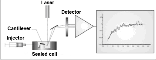
The schematic diagram of the device
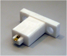
Cantilevers are mounted in the special holder
Convenient software
The BioScan Atomic Scales system is controlled by the FemtoScan VideoSense program. The upper part of the program window shows the image of the sensor cell taken by the camera, the signal arriving at the detector and the processed image, in which the reflected rays are shown in different colors. The following graphs are constructed depending on the deviation of each console from time.

Advanced Technologies is a member of the Skolkovo Innovation Center